
Overview of Human Germ Cell Developmental Stages and Critical Windows
By Meeri Kim, PhD
|
Humans have two primary types of cells. Germ cells are those involved in reproduction and include the well-known male and female gametes – egg and sperm, respectively – as well as their developmental precursors. By definition, somatic cells make up the remainder of an organism’s body such as skin, bones, organs, and blood.
Germ cells are essential to ensure the correct passage of DNA from one generation to the next. They represent the molecular phase of human development, with their own complex life cycle. It begins with the setting aside of primordial germ cells in the early embryo (specification) and ends with sperm and egg fusing together to begin a new life (fertilization). Throughout this life cycle, which generally lasts up to 40 years, there are several time-sensitive intervals during which exposures or events can disrupt development. These so-called critical windows leave the embryo vulnerable to adverse and irreversible epigenetic effects. What follows is a brief outline of the stages of germ cell development with critical windows highlighted. |
During the germline lifecycle there are several time-sensitive intervals during which exposures or events can disrupt development.
|
|
1. Specification
The germ cell lineage is one of the first to form in the human embryo. During early embryonic development, a small portion of cells are set aside through inductive signaling to later become germ cells. This process is termed specification, and at this early stage, they are called primordial germ cells (PGCs) and are located outside the embryo. In humans, PGCs are formed in the second week of development. In this period, they are thought to be epigenetically similar to the somatic cells around them. |
Human primordial germ cells are formed in the second week of embryonic development.
|
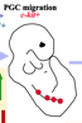
2. Migration
Once specified, PGCs begin to migrate from the yolk sac towards their final destination – the gonads. During this migration, which occurs during the third to fifth week of development (5-7 weeks of pregnancy), the cells begin to multiply and mature. Their epigenetic marks are remodeled and soon appear distinct from those of the surrounding somatic cells. Finally, the PGCs colonize the gonads. This migration period is a key critical window. At this stage, when major changes are occurring in DNA methylation and other aspects of chromatin marking, environmental exposures can have long-term repercussions. The embryo at this time is therefore very sensitive to enduring epigenetic modifications. |
Germ cells must actively migrate to the gonads. During migration their epigenetic marks are remodeled and sensitive to enduring modifications.
|
3. Germline During Fetal Development
Once the PGCs have reached the gonads, they begin to develop differently according to their sex. At this point, sexual differentiation splits the germ cell lifecycle into distinct branches for a female versus male embryo. As proliferation continues, old epigenetic information – even some residual epigenetic marks inherited from parents in the form of imprinted genes – are removed. For both sexes at this stage, the DNA of the germline lacks the protective effects of methylation, leaving it very sensitive to environmental damage. Because genes are imprinted differently depending on whether they are of maternal or paternal origin, this erasure is essential to ensure that the epigenetic marks in the PGCs are reset and appropriately reflect the sex of the developing embryo. Note that the imprinted genes of the somatic cells are not demethylated. The timing of this process happens also depends on sex. In females, establishment of the epigenetic female imprint occurs post-embryonically and after the first stage of meiosis is complete. However, in males, establishment of the male imprint begins shortly after sex determination but before meiosis. |
Once in the gonads the germ cells are demethylated, leaving them sensitive to environmental damage. New epigenetic imprints are then placed on a subset of genes, depending on whether the embryo is male or female.
|
4. Oogenesis The germline of the female is formed during embryonic development prior to birth, meaning that this period is undoubtedly a critical window for future generations. The process of oogenesis, or the production of female gametes, begins when the PGCs migrate into the developing ovaries to form small nests of immature germ cells called oogonia. The diploid oogonia continue to undergo mitosis until middle fetal life, when all cell divisions have been completed. Their number peaks at approximately 6 to 7 million at 20 weeks of gestation. Then, a subset undergo programmed cell death. The remaining cells – known as primary oocytes – arrest in prophase-I of meiosis and remain dormant until just prior to ovulation, after puberty. The number of primary oocytes gradually declines to about 2 to 2.5 million at birth. |
The germline of the female is formed during embryonic development prior to birth, meaning that this period is undoubtedly a critical window for future generations.
|
5. Spermatogenesis
In males, the PGCs migrate into the developing testes and undergo further proliferation until 16 to 18 weeks of gestation. Then a number go through programmed cell death, similar to what happens in females, before arresting in mitosis within the seminiferous tubules. These germ cells are diploid, mitotically active, self-renewing spermatogonia. During adolescence, the spermatogonia begin to multiply again and finally undergo meiosis to form sperm. In males, sperm production is a continuously ongoing process from adolescence onward. The final touches on the epigenetic profiles in sperm are made as they go through the last stages of maturation. This means that they could be vulnerable to environmental insults over the entire course of a male’s lifetime. On the other hand, sperm produced months after a harmful exposure may remain unharmed, as long as there has not been extensive damage to the germline stem cells. |
In males, sperm production is a continuously ongoing process from adolescence onward. The final touches on the epigenetic profiles in sperm are made as they go through the last stages of maturation.
|
|
6. Germline through Childhood
After birth in the female, the germ cells are kept in a suspended state – meiotic arrest in prophase I – for many years, up to decades. They remain more or less unchanged from early embryogenesis until they are used for ovulation in the adult. During this period, any damage due to adverse environmental exposure could accumulate. Also, the total number of primary oocytes continues to decline during childhood until they number at about 400,000 right before puberty. Similarly, the male spermatogonia lie in wait until puberty and do not multiply. But the difference is that these cells have not lost their ability to multiply, and begin to do so again at puberty. |
During this period, any damage due to adverse environmental exposure could accumulate.
|
|
7. Germline in Puberty
In both sexes, mature germ cells are not fully formed until puberty. In females, through a process called folliculogenesis, the primary oocyte completes its first meiotic cycle and extrudes the first polar body at ovulation. In males, the spermatogonia activate and start to proliferate. Differentiation is yet another critical window, where DNA is re-packed and sensitive to environmental factors. Morphologically mature spermatozoa are produced by spermatogonia meiosis in the seminiferous tubules of the testes – a process that takes about 48 days. A healthy human male produces somewhere between 45 and 207 million spermatozoa per day that are then released into the central lumen of the seminiferous tubule. |
A healthy human male produces somewhere between 45 and 207 million spermatozoa per day that are then released into the central lumen of the seminiferous tubule.
|
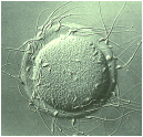
8. Conception
After ovulation, the egg enters the female reproductive tract where fertilization can take place by a sperm. Once a single sperm penetrates through to the egg, it becomes impermeable to additional sperm. The male DNA becomes decondensed and rapidly demethylated. After fertilization, the fused sperm and egg is called a zygote. The zygote contains a full diploid set of DNA – half comes from the haploid egg, and the other from the haploid sperm. |
Once a single sperm penetrates through to the egg, it becomes impermeable to additional sperm. The male DNA becomes decondensed and rapidly demethylated.
|
|
9. Pre-Implantation
For the next few days, the zygote travels down the Fallopian tube and divides mitotically. First, it becomes a solid mass of cells called a morula. This stage is followed by the formation of a cavity within the ball. The blastocyst, as it is now known, contains an inner group of cells – to become the embryo – and an outer layer that will form protective membranes. Embryos during the morula/early blastocyst stage are considered to bear much lower levels of epigenetic marks than zygotes. The period of blastocyst formation includes extensive epigenetic reprogramming of the parental genomes, including genome-wide DNA demethylation. Once the blastocyst reaches the uterus, around week 4 of gestation, it implants itself tightly to the uterine wall and begins to receive nourishment from the mother’s blood. Around the time of implantation, the reprogramming process is largely complete. |
The period of blastocyst formation includes extensive epigenetic reprogramming of the parental genomes, including genome-wide DNA demethylation.
|